Tag: Medical Knowledge

  • Armpit Lump Causes: 4 Types and Warning Signs to Know

    Armpit Lump Causes: 4 Types and Warning Signs to Know

    SummaryMistaking an armpit lump for a simple pimple and squeezing it or ignoring it can lead to much higher treatment costs down the road. Understanding the main armpit lump causes can help you catch problems early. Explore the 4 possible causes — sebaceous cysts, lymphadenitis, accessory breast tissue, and lipomas — along with their warning signs and proper treatment options.

    While massaging your lymph nodes,
    have you ever touched your armpit
    and felt a hard lump underneath?

    Brushing it off as "just a big pimple"
    can sometimes lead to spiraling medical bills later.

    That’s because an armpit lump isn’t always a simple pimple —
    it can be a warning sign of a condition that needs treatment.
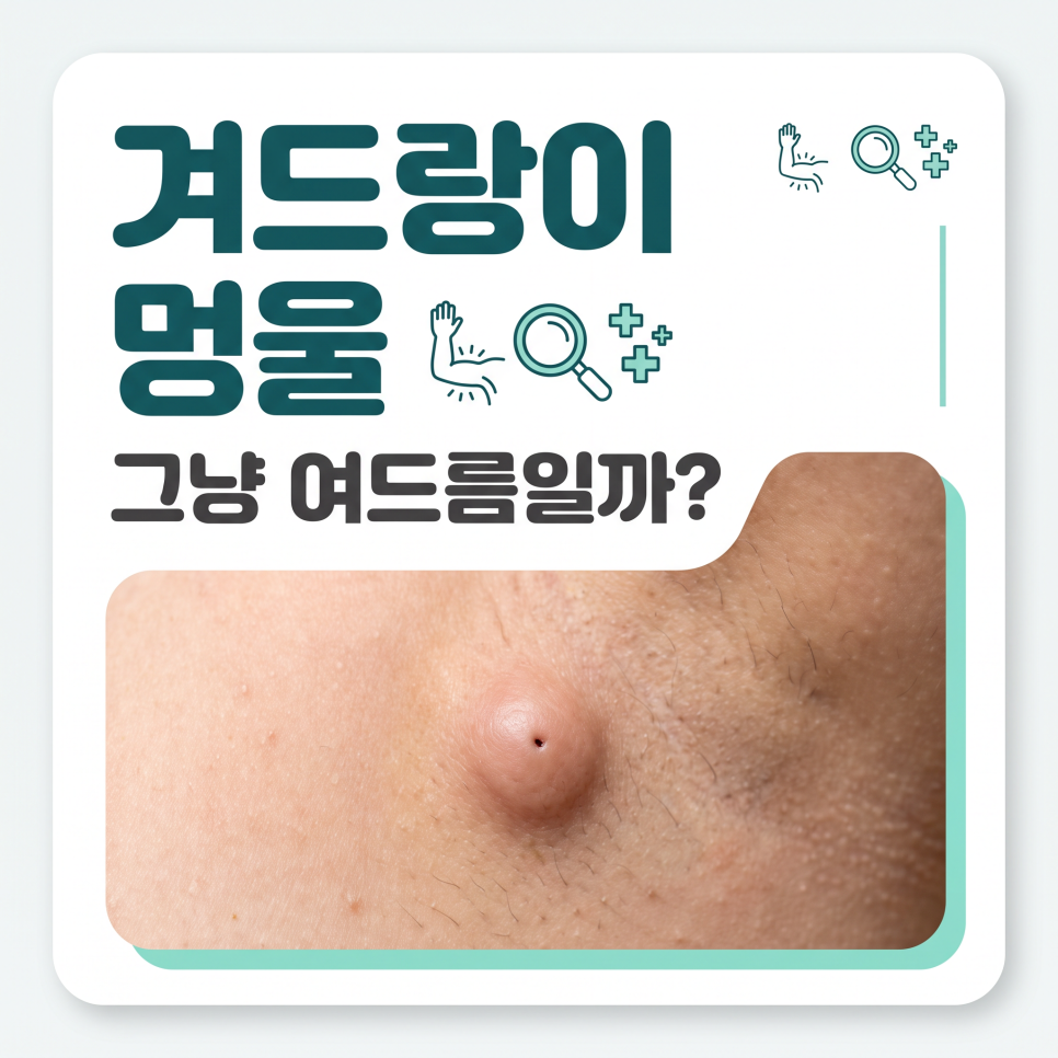
    armpit lump causes

    Key Takeaways
    An armpit lump can have many different causes, including sebaceous cysts, lymphadenitis, accessory breast tissue, and lipomas.
    It’s safest to monitor for pain and size changes and get an ultrasound exam for an accurate diagnosis.
    Some cases resolve on their own depending on the cause, but leaving it unchecked can increase the scope and cost of treatment.

    What Are the Main Armpit Lump Causes?

    To answer the question directly, armpit lump causes generally fall into one of four main categories.
    An armpit lump is a mass that forms in the skin or the tissue beneath it, and the right treatment depends entirely on its underlying cause.

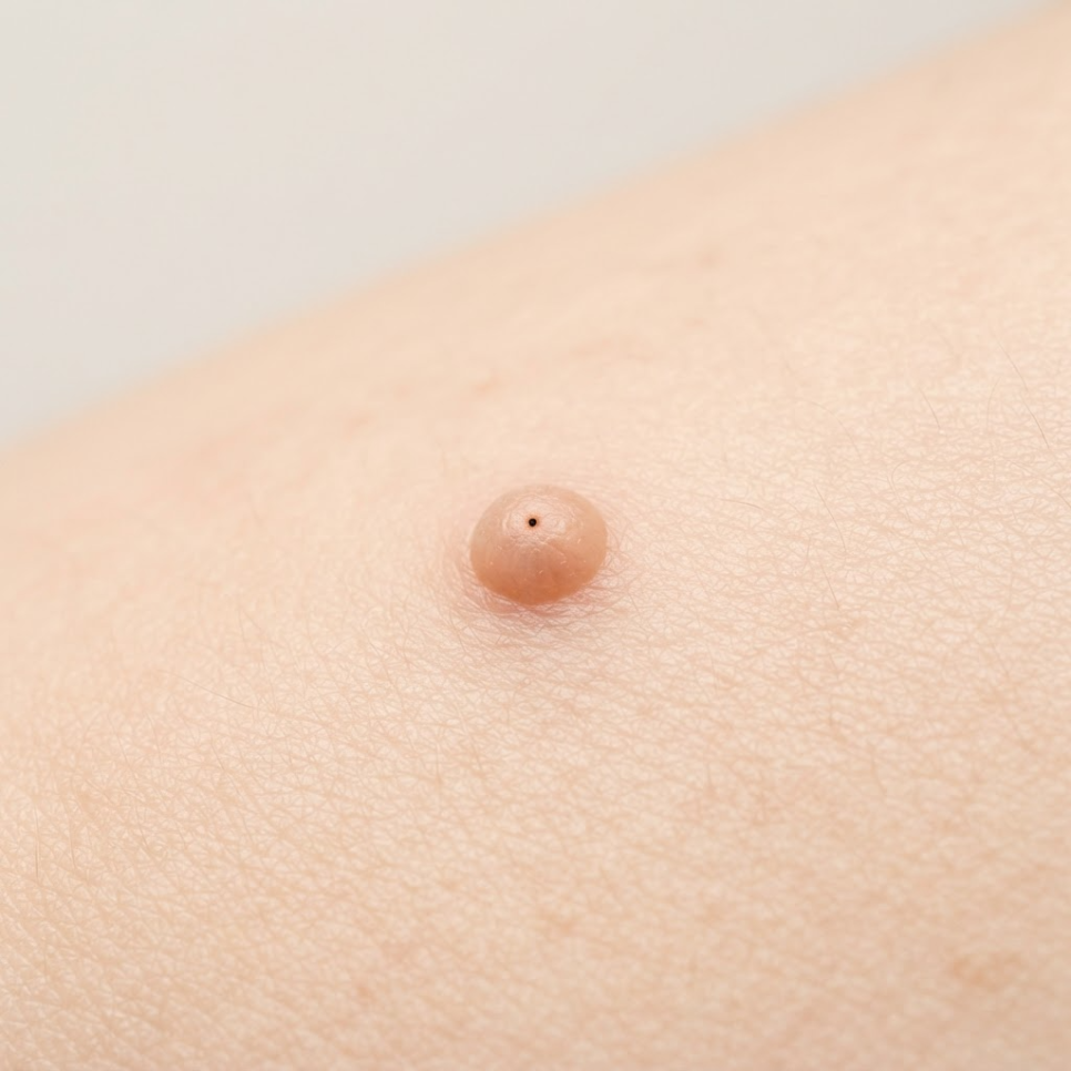
    A common cause is an epidermal cyst (sebaceous cyst), which forms when a hair follicle or sebaceous material builds up under the skin.
    According to Asan Medical Center’s disease encyclopedia, epidermal cysts typically grow slowly to about 1 to 5 cm and sometimes show a small central opening (punctum).
    Squeezing the cyst can release foul-smelling discharge, but doing so carelessly can worsen inflammation, so caution is needed.
    armpit lump related image 2

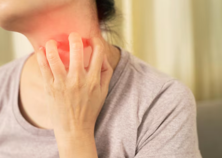
    The second cause is lymphadenitis (swollen lymph nodes), where the lymph nodes enlarge as part of an immune response.
    When the body reacts to a cold, weakened immunity, or internal inflammation, the lymph nodes swell and can be felt as a firm lump.
    Asan Medical Center describes lymphadenitis as "an immune response in which the lymph nodes enlarge due to infection or, less commonly, malignant tumors."
    armpit lump related image 3

    The third cause is accessory breast tissue, which occurs when mammary gland tissue remains in the armpit area.
    It develops when the "milk line" tissue running from the chest to the armpit during fetal development doesn’t fully regress, and its size and pain level change with the menstrual cycle and hormonal shifts.
    Finally, a soft, slow-growing, painless lipoma is another common cause.

    Type Characteristics Pain
    Epidermal/Sebaceous Cyst Close to skin surface, may discharge fluid Usually none
    Lymphadenitis Caused by immune response Painful when pressed
    Accessory Breast Tissue Size fluctuates with menstrual cycle Cyclical pain
    Lipoma Soft, slow-growing None
    Checklist
    ✅ Epidermal/Sebaceous cyst: Feels soft just beneath the skin and may discharge fluid
    ✅ Lymphadenitis: Occurs from an immune response and causes pain when pressed
    ✅ Accessory breast tissue: Residual mammary tissue that changes in pain and size with the menstrual cycle
    ✅ Lipoma: A soft, painless fatty lump that grows slowly

    Is a Painless Armpit Lump Safe to Ignore?

    No. In fact, a hard, painless lump deserves even more attention.
    A warning sign isn’t judged by how much it hurts, but by changes in the lump’s size, firmness, and whether it’s fixed in place.

    Warning Signs
    ⚠️ When it feels completely painless, hard, and fixed in place when touched
    ⚠️ When it gradually grows larger over time and nearby skin changes appear
    ⚠️ When fever, redness, or swelling around the armpit worsens
    • A normal lymph node is usually less than 1 cm in diameter, small, and moves freely.
    • When inflammation progresses, however, the lymph node adheres to surrounding tissue and becomes difficult to move.
    • If the size, firmness, or mobility changes, further testing should be considered.

    Lymph node enlargement occurs when the lymph nodes are infected or inflamed, or in cases involving a pus-filled abscess or cancer.

    The Korea Disease Control and Prevention Agency explains this through its National Health Information Portal, noting that localized lymph node enlargement accounts for 70 to 80 percent of all cases.
    Among these, swelling in the neck’s cervical lymph nodes is common, with the armpit and groin also being frequent sites.
    armpit lump related image 4

    A painless lump is more likely to be a chronic cyst or lipoma, but in rare cases further testing is necessary.
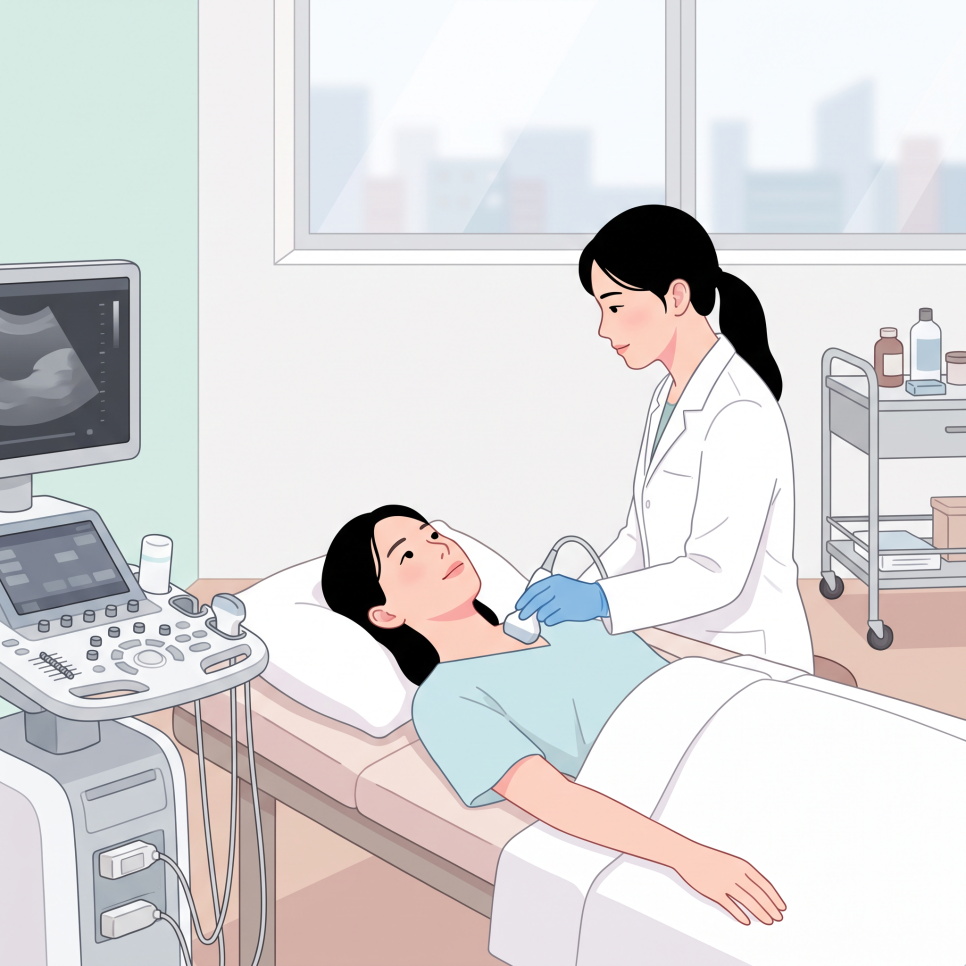
    That’s why confirming the diagnosis with an ultrasound exam is a safer choice than self-diagnosis.


    How Does Treatment Differ by Cause?

    Knowing the exact cause is key to reducing recurrence and complications.
    Treatment for an armpit lump ranges from simple monitoring to surgical removal, depending entirely on the underlying condition.

    1. Lymphadenitis: May improve with adequate rest and immune care alone; anti-inflammatory pain relievers or antibiotics may be prescribed if needed.
    2. armpit lump related image 5

    3. Sebaceous cyst (epidermal cyst): Avoid squeezing it by hand; having it surgically removed under local anesthesia, including the cyst wall, can lower the risk of recurrence.
    4. armpit lump related image 6

    5. Accessory breast tissue: Since hormones play a major role, periodic monitoring is the standard approach, and surgical removal of the glandular tissue may be considered if pain or discomfort significantly affects daily life.
    Outcome
    Identifying armpit lump causes accurately and managing them appropriately may help some cases avoid major surgery, depending on the underlying condition.
    However, we recommend discussing the diagnosis and treatment plan with a medical professional before making any decisions.

    If you’re concerned about a lump or wound on your skin — not just in the armpit — seeing a specialist is important.
    Burn Treatment Specialists – [Ansan Open Surgery Hospital] Homepage has more information on surgical care.


    Frequently Asked Questions

    1. Is it okay to keep touching an armpit lump to check on it?
    Answer: Bacteria on your hands can cause a secondary infection or worsen inflammation, so it’s best to avoid pressing on or repeatedly touching the lump.

    2. If an armpit lump doesn’t hurt, is it fine to just leave it alone?
    Answer: Even without pain, it could be a chronic cyst, lipoma, or another condition, so it’s safer to monitor any size changes and confirm the diagnosis with an ultrasound exam.

    3. Why does my armpit lump only hurt during my period?

    Answer: It’s likely accessory breast tissue responding to changes in female hormones. This tissue tends to temporarily swell and become painful in sync with the menstrual cycle, then subside afterward.

    4. Which specialist should I see for an armpit lump?
    Answer: Visiting a breast surgeon, general surgeon, or dermatologist allows for an accurate diagnosis using tools like ultrasound, along with guidance tailored to the cause.

    armpit lump related image 7


    References

    · Korea Disease Control and Prevention Agency, "Lymph Node Enlargement," National Health Information Portal. Link
    · Asan Medical Center, "Lymphadenitis," Disease Encyclopedia. Link
    · Asan Medical Center, "Epidermal Cyst," Disease Encyclopedia. Link
    · Health Kyunghyang, "[Accessory Breast Story] Accessory Breast Tissue Is a Real Medical Condition," 2023. Link

    This article is provided for general health information purposes only and is not a substitute for individual diagnosis or treatment. If you have symptoms, please consult a medical professional.
  • Lipoma Size 5cm or Larger: 3 Signs Surgery May Be Needed

    Lipoma Size 5cm or Larger: 3 Signs Surgery May Be Needed

    SummaryThis article summarizes medical guidance on whether a lipoma size of 5cm or larger requires surgery, along with how lipomas are diagnosed and managed.

    Finding a soft, movable lump under your skin can be alarming at first.
    Most of the time there’s no pain or color change, so it’s easy to brush off.

    But once you notice it slowly growing larger over time, the concern becomes harder to ignore.
    You may start wondering whether surgery is necessary, and just how large a lipoma size has to be before it becomes a real concern.

    This is actually a question that comes up often in clinical practice.

    This article outlines the surgical criteria based on changes in lipoma size, along with accurate diagnostic methods.
    Let’s look at what standards doctors use to consider excision when a lipoma grows beyond 5 centimeters or becomes painful, and how to manage it safely without unnecessary worry.

    lipoma size

    What Is a Lipoma and Why Does It Form?

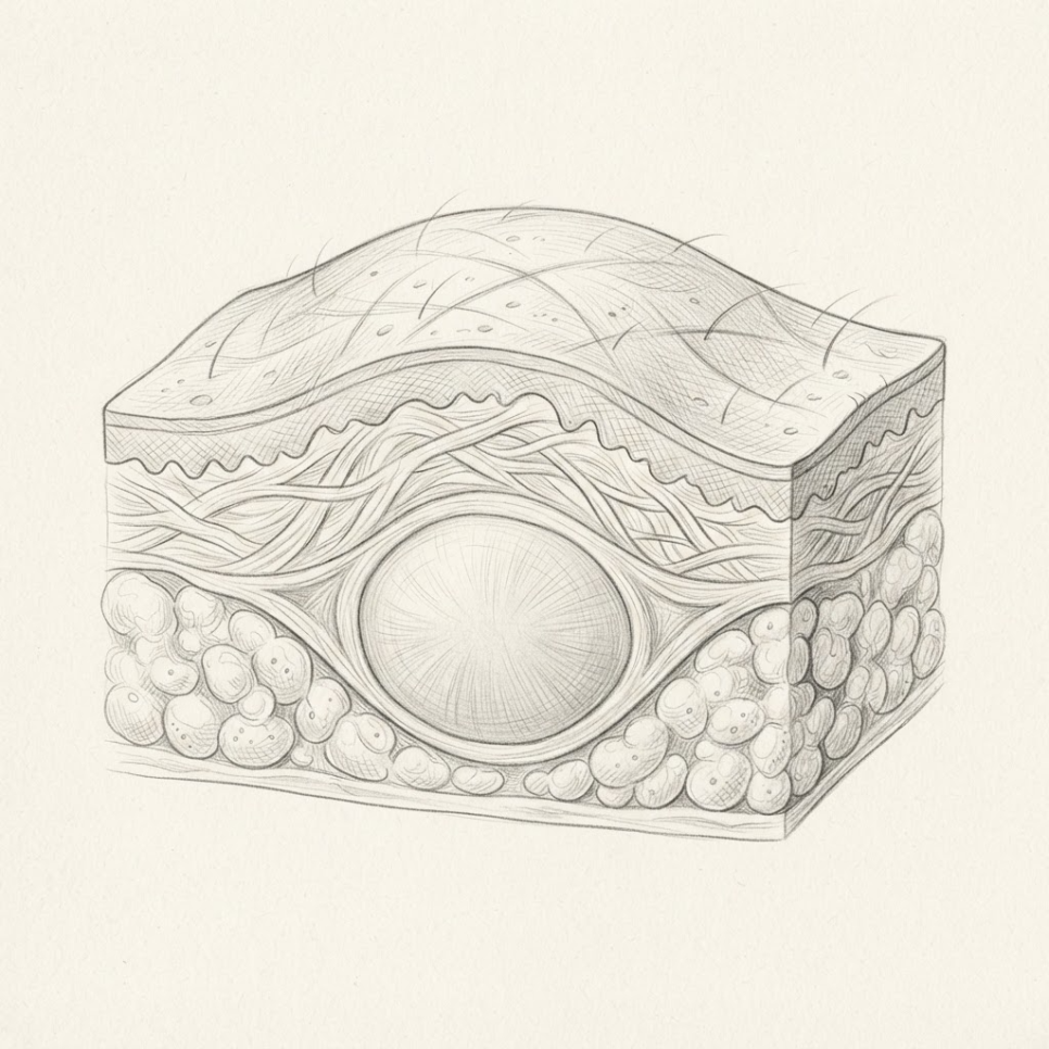
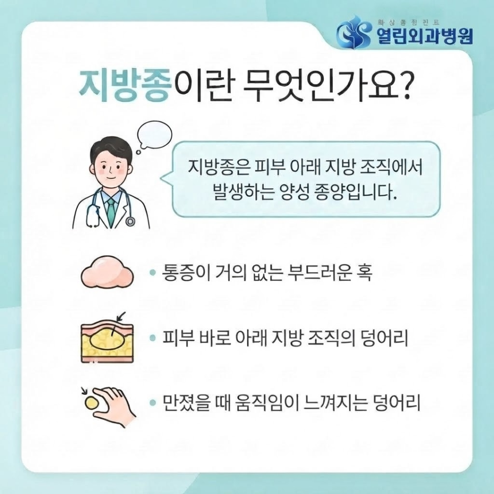
    A lipoma is a common benign tumor that forms when fat cells cluster together and grow within the subcutaneous fat tissue.
    It’s common enough that it may develop in adults of any age.

    Because it’s usually painless, it’s easy to dismiss at first.
    But it’s completely natural to feel a jolt of worry when you discover an unfamiliar lump on your body.

    That’s why doctors often emphasize the importance of distinguishing a lipoma from liposarcoma, a malignant tumor it can sometimes be confused with.

    lipoma size related image 2

    Key Point
    A lipoma is typically painless and moves easily when pressed with a finger. If a lump feels hard and stays fixed in place, other conditions should be ruled out.

    Lipoma Size: How Large Can It Grow?

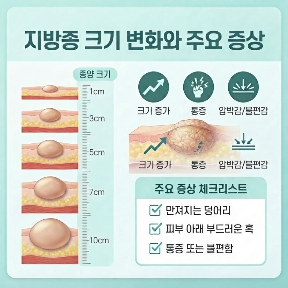
    Lipoma size typically ranges from about 1 centimeter to several centimeters.
    Some grow slowly over time, while others remain unchanged for years.

    lipoma size related image 3

    In rare cases, a lipoma can grow beyond 10 centimeters, becoming what’s known as a giant lipoma.
    As it grows larger, it can press on nearby nerves or muscles.

    This may bring on pain, tingling, or general discomfort.
    The change in appearance can also be a source of stress.

    Checklist
    ✅ Has the lump grown noticeably larger over the past few months?
    ✅ Does it feel hard and fixed in one place when touched?
    ✅ Do you feel pressure or pain during everyday activities?
    ✅ Is the surface irregular or the border unclear?

    lipoma size related image 4


    Does Lipoma Size Determine Whether You Need Surgery?

    Not every lipoma needs to be surgically removed.
    If it’s small and causes no particular discomfort, simply monitoring it over time is often enough.

    However, excision may be considered when a lipoma meets specific medical criteria.
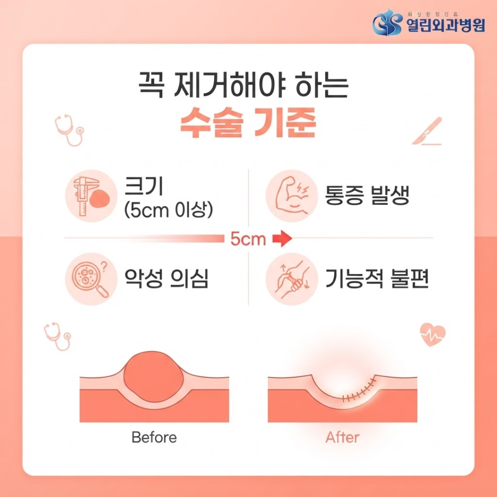
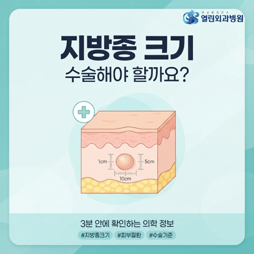
    According to disease information from Asan Medical Center, treatment is generally considered when the lipoma is 5 centimeters or larger, or when it causes pain or grows at an abnormal rate.

    lipoma size related image 5

    Category Monitor (Watchful Waiting) Consider Surgery
    Size Under 5cm, little to no change 5cm or larger, or growing rapidly
    Symptoms No pain or pressure Pain, tingling, or functional impairment
    Imaging findings Typical benign appearance Findings suggestive of malignancy, such as liposarcoma

    Beyond cosmetic concerns, these functional symptoms are what typically determine whether surgery is warranted.

    Outcomes
    Excision performed based on clear medical criteria may help lower the risk of recurrence and may also help ease everyday discomfort such as pressure or pain. Outcomes can vary from person to person.

    How Are Lipomas Diagnosed and Managed?

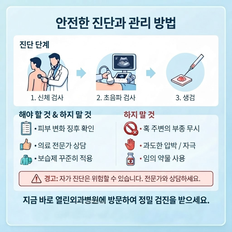
    Lipoma diagnosis is generally made through a physical exam combined with an ultrasound.
    Visual inspection or palpation alone can’t reliably determine the exact depth and characteristics of the mass.

    lipoma size related image 6

    Medical information from Seoul National University Hospital notes that when differentiation from liposarcoma is needed, CT, MRI, or a tissue biopsy may be performed as well.
    According to the Korea Disease Control and Prevention Agency’s National Health Information Portal, ultrasound-guided biopsy allows the lesion’s location to be confirmed in real time while collecting tissue for an accurate diagnosis.

    Relying on online reviews or unverified home remedies can sometimes lead to a delayed diagnosis.
    Getting an objective evaluation at a nearby medical facility is generally recommended.

    If you need an accurate diagnosis and consultation, you’re welcome to check the care information at Ansan Yeorin Surgery Hospital, a burn treatment specialist – Homepage.

    lipoma size related image 7

    Caution
    Do not attempt to press on, squeeze, or forcibly remove the lump yourself based on self-diagnosis — this can lead to inflammation or infection and should be strictly avoided.

    Frequently Asked Questions

    1. Can a lipoma go away on its own?
    Answer: It’s rare for a lipoma to shrink or disappear naturally — it typically stays the same size or grows gradually over time.

    2. How painful is the surgery, and how long is recovery?
    Answer: Most cases involve a simple excision under local anesthesia, and while it varies by size, patients typically return to daily activities relatively quickly.

    3. Is this condition likely to come back after treatment?
    Answer: Once fully excised, recurrence at the same site is relatively uncommon, though a new lipoma may still develop elsewhere.

    4. Should I still see a doctor if the lipoma is small?
    Answer: Even though it’s a benign tumor, an early evaluation is recommended to rule out other conditions such as liposarcoma.

    5. How is a lipoma different from a cyst?
    Answer: Because texture, location, and ultrasound findings differ between the two, a proper exam is needed rather than relying on visual assessment alone.

    lipoma size related image 8

    lipoma size related image 9

    lipoma size related image 10

    lipoma size related image 11


    References

    · Asan Medical Center, “Lipoma,” Disease Encyclopedia, Asan Medical Center. View source
    · Seoul National University Hospital, “Lipoma,” Medical Information, Seoul National University Hospital. View source
    · Korea Disease Control and Prevention Agency, “Ultrasound-Guided Biopsy,” National Health Information Portal, Korea Disease Control and Prevention Agency. View source


    This article is provided for general health information purposes only and is not a substitute for individual diagnosis or treatment. If you have symptoms, please consult a medical professional.
  • Flap Necrosis: 3 Causes After Free Flap Surgery (Smoking & Pressure Warning)

    Flap Necrosis: 3 Causes After Free Flap Surgery (Smoking & Pressure Warning)

    SummarySmoking and pressure on the graft site are two of the leading causes of flap necrosis after free flap surgery. Here’s a rundown of reconstructive plastic surgery information — from the basics of vascular anastomosis to a post-surgery care checklist.

    What if a flap that just went through surgery suddenly turned dark red or purple?
    Tissue transferred through free flap surgery is at risk of flap necrosis the moment its blood supply is interrupted.

    Smoking and pressure from improper positioning are two of the most common causes.
    Today, we’ll walk through how free flap surgery works and how to help prevent flap necrosis after the procedure.

    flap necrosis

    Key Points
    ✔ Free flap surgery is a microsurgical reconstruction procedure that transplants tissue along with its blood vessels.
    ✔ Inadequate post-surgery care can lead to flap necrosis.
    ✔ Smoking and pressure are two of the leading risk factors for flap necrosis.

    What Is Free Flap Surgery?

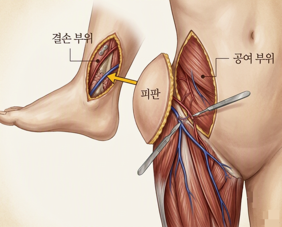
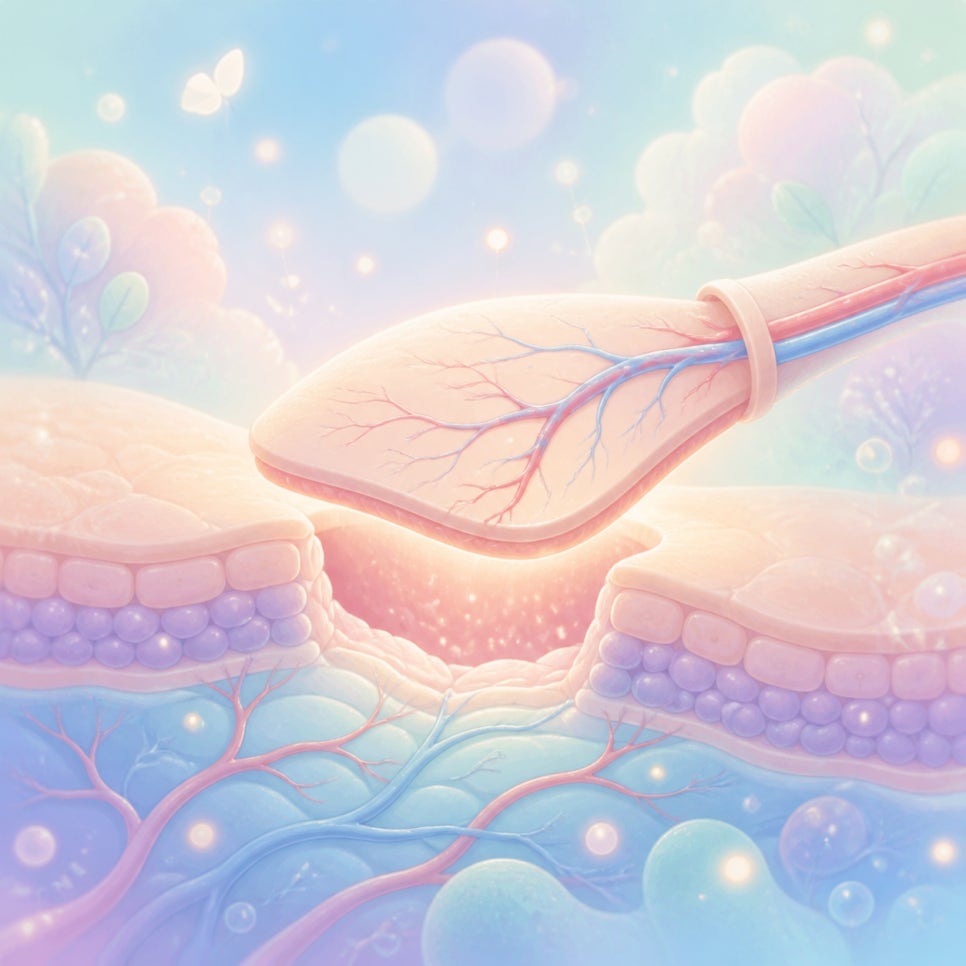
    To put it simply, free flap surgery is a microsurgical reconstruction procedure that removes a whole section of a patient’s own tissue — including its blood vessels — and transplants it to a damaged area.

    flap necrosis illustration 2

    In some cases, a simple skin graft isn’t enough for full recovery.
    This is especially true when the tissue damage is deep or blood circulation to the area is compromised.

    In these situations, surgeons take skin, fat, and even arteries and veins together from a donor site such as the thigh, back, or abdomen.
    But that’s not the end of the process.

    flap necrosis illustration 3

    The tiny blood vessels in the removed tissue must be connected, one by one, to the blood vessels at the recipient site under a microscope.
    This vascular anastomosis process is the core technique of free flap surgery.

    That’s because tissue needs a continuous supply of oxygen and nutrients to survive.
    According to related research, this microvascular connection is considered a critical factor in determining whether the surgery succeeds.

    flap necrosis illustration 4


    Why Does Flap Necrosis Happen? The Effects of Smoking and Pressure

    In short, flap necrosis occurs when the oxygen and nutrient supply to transplanted tissue is cut off, causing the cells to die.

    Nicotine in cigarettes causes blood vessels to constrict.
    Even a single cigarette can narrow the microvasculature.
    That’s why smoking after surgery is considered one of the leading risk factors for flap necrosis.

    Pressure has a similar effect.
    Something as simple as an unconscious sleeping position can cut off blood flow to the grafted area.
    When a blood vessel is compressed, blood clots can form more easily, which can ultimately lead to tissue necrosis.

    ⚠ Precautions
    · Maintain a position that keeps pressure off the surgical site at all times.
    · Avoid smoking, since it constricts blood vessels.
    · Keep warm, as a drop in body temperature can slow blood circulation.
    Risk Factor Effect on the Flap
    Smoking Vasoconstriction, reduced microcirculation
    Pressure/positioning Vessel compression, increased risk of clot formation
    Hypothermia Reduced blood circulation

    An international meta-analysis found that smoking significantly increased the rates of hematoma and overall complications after head and neck cancer reconstruction surgery.
    Another study reported that free flap perfusion tended to be lower in smokers than in non-smokers.

    That said, having a smoking history doesn’t automatically mean flap necrosis will occur.
    Still, since the risk is elevated, extra caution is warranted.

    flap necrosis illustration 5


    When and How Is Free Flap Surgery Performed?

    In short, free flap surgery is used when extensive tissue loss needs to be structurally restored.
    It’s commonly used to fill defects left after severe burns, head and neck cancer surgery, or breast cancer surgery.
    It’s also widely applied for serious skin damage caused by traffic accidents or diabetic foot conditions.

    Checklist — Steps in Free Flap Surgery
    ✅ Step 1: Measure the defect and select the donor site (where tissue will be harvested)
    ✅ Step 2: Harvest the flap, including its blood vessels, from the donor site
    ✅ Step 3: Perform vascular anastomosis (vessel connection) using a surgical microscope
    ✅ Step 4: Close the recipient site and the donor site

    Because this microsurgical technique involves connecting blood vessels with sutures thinner than a human hair, the procedure takes a long time and requires a high degree of precision.


    What Should You Manage After Surgery?

    To put it simply, the key to post-surgery care is keeping the transplanted blood vessels open to prevent flap necrosis.

    flap necrosis illustration 6

    After surgery, blood flow to the transplanted tissue must be monitored continuously.
    If a vessel becomes blocked or a clot forms, the transplanted tissue is unlikely to survive.

    Close monitoring takes place during the first several days.
    Once the tissue has stabilized, patients gradually move into a more routine recovery phase.

    When recovery is managed well,
    it can help support stable flap survival and normal blood flow, and may also help lower the risk of tissue necrosis.

    If you’d like to learn more about tissue defects caused by burns and reconstructive treatment options, visiting Burn Treatment, [Ansan Open Surgery Hospital] Home may also be helpful.

    flap necrosis illustration 7


    Frequently Asked Questions

    1. Can tissue from another person be transplanted instead?
    No — to prevent immune rejection, the general rule is to use only the patient’s own tissue.

    2. Is the donor site (where tissue is harvested) affected afterward?
    Surgeons choose a site that has minimal impact on bodily function and where scarring is less visible, and the donor site is closed with careful surgical technique as well.

    3. How long does recovery take after surgery?
    Hospital stays typically last about one to two weeks, but several months of follow-up monitoring are usually needed before the graft site stabilizes and the patient returns to normal daily activities.

    4. Can the skin color or texture differ from the surrounding area?
    Depending on the donor site used, there can be differences in texture or color, and additional cosmetic procedures may be used to correct this later if needed.

    5. Can smokers undergo free flap surgery?
    Smoking doesn’t necessarily rule out surgery, but because it raises the risk of flap necrosis, quitting smoking before and after surgery is strongly recommended.


    References

    · Asan Medical Center, “Tissue Reconstruction Through Flap Surgery,” Disease Encyclopedia, Asan Medical Center. View source
    · Chang et al., “The impact of smoking on surgical complications after head and neck reconstructive surgery with a free vascularised tissue flap: a systematic review and meta-analysis,” Journal of Plastic, Reconstructive & Aesthetic Surgery (ScienceDirect), 2020. View source
    · “Smoking and microvascular free flap perfusion in head and neck reconstruction: radial free forearm flaps and anterolateral thigh flaps,” Scientific Reports (Nature), 2022. View source
    · Liu et al., “Analysis of the Risk Factors for Free Flap Necrosis in Soft Tissue Reconstruction of the Lower Limbs,” Orthopaedic Surgery (Wiley), 2023. View source

    This article is intended for general health information purposes only and is not a substitute for individual diagnosis or treatment. If you have symptoms, please consult a medical professional.