Tag: Health Guide

  • Armpit Lump Causes: 4 Types and Warning Signs to Know

    Armpit Lump Causes: 4 Types and Warning Signs to Know

    SummaryMistaking an armpit lump for a simple pimple and squeezing it or ignoring it can lead to much higher treatment costs down the road. Understanding the main armpit lump causes can help you catch problems early. Explore the 4 possible causes — sebaceous cysts, lymphadenitis, accessory breast tissue, and lipomas — along with their warning signs and proper treatment options.

    While massaging your lymph nodes,
    have you ever touched your armpit
    and felt a hard lump underneath?

    Brushing it off as "just a big pimple"
    can sometimes lead to spiraling medical bills later.

    That’s because an armpit lump isn’t always a simple pimple —
    it can be a warning sign of a condition that needs treatment.
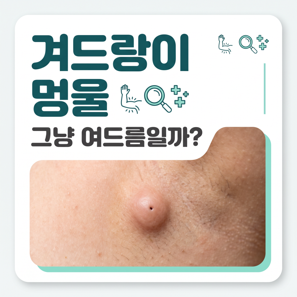
    armpit lump causes

    Key Takeaways
    An armpit lump can have many different causes, including sebaceous cysts, lymphadenitis, accessory breast tissue, and lipomas.
    It’s safest to monitor for pain and size changes and get an ultrasound exam for an accurate diagnosis.
    Some cases resolve on their own depending on the cause, but leaving it unchecked can increase the scope and cost of treatment.

    What Are the Main Armpit Lump Causes?

    To answer the question directly, armpit lump causes generally fall into one of four main categories.
    An armpit lump is a mass that forms in the skin or the tissue beneath it, and the right treatment depends entirely on its underlying cause.

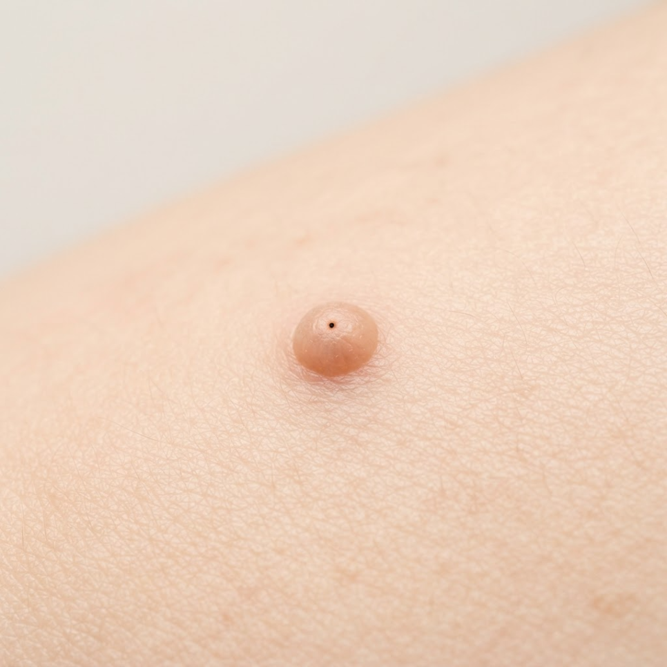
    A common cause is an epidermal cyst (sebaceous cyst), which forms when a hair follicle or sebaceous material builds up under the skin.
    According to Asan Medical Center’s disease encyclopedia, epidermal cysts typically grow slowly to about 1 to 5 cm and sometimes show a small central opening (punctum).
    Squeezing the cyst can release foul-smelling discharge, but doing so carelessly can worsen inflammation, so caution is needed.
    armpit lump related image 2

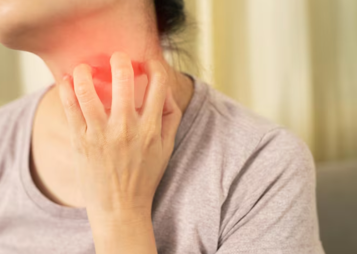
    The second cause is lymphadenitis (swollen lymph nodes), where the lymph nodes enlarge as part of an immune response.
    When the body reacts to a cold, weakened immunity, or internal inflammation, the lymph nodes swell and can be felt as a firm lump.
    Asan Medical Center describes lymphadenitis as "an immune response in which the lymph nodes enlarge due to infection or, less commonly, malignant tumors."
    armpit lump related image 3

    The third cause is accessory breast tissue, which occurs when mammary gland tissue remains in the armpit area.
    It develops when the "milk line" tissue running from the chest to the armpit during fetal development doesn’t fully regress, and its size and pain level change with the menstrual cycle and hormonal shifts.
    Finally, a soft, slow-growing, painless lipoma is another common cause.

    Type Characteristics Pain
    Epidermal/Sebaceous Cyst Close to skin surface, may discharge fluid Usually none
    Lymphadenitis Caused by immune response Painful when pressed
    Accessory Breast Tissue Size fluctuates with menstrual cycle Cyclical pain
    Lipoma Soft, slow-growing None
    Checklist
    ✅ Epidermal/Sebaceous cyst: Feels soft just beneath the skin and may discharge fluid
    ✅ Lymphadenitis: Occurs from an immune response and causes pain when pressed
    ✅ Accessory breast tissue: Residual mammary tissue that changes in pain and size with the menstrual cycle
    ✅ Lipoma: A soft, painless fatty lump that grows slowly

    Is a Painless Armpit Lump Safe to Ignore?

    No. In fact, a hard, painless lump deserves even more attention.
    A warning sign isn’t judged by how much it hurts, but by changes in the lump’s size, firmness, and whether it’s fixed in place.

    Warning Signs
    ⚠️ When it feels completely painless, hard, and fixed in place when touched
    ⚠️ When it gradually grows larger over time and nearby skin changes appear
    ⚠️ When fever, redness, or swelling around the armpit worsens
    • A normal lymph node is usually less than 1 cm in diameter, small, and moves freely.
    • When inflammation progresses, however, the lymph node adheres to surrounding tissue and becomes difficult to move.
    • If the size, firmness, or mobility changes, further testing should be considered.

    Lymph node enlargement occurs when the lymph nodes are infected or inflamed, or in cases involving a pus-filled abscess or cancer.

    The Korea Disease Control and Prevention Agency explains this through its National Health Information Portal, noting that localized lymph node enlargement accounts for 70 to 80 percent of all cases.
    Among these, swelling in the neck’s cervical lymph nodes is common, with the armpit and groin also being frequent sites.
    armpit lump related image 4

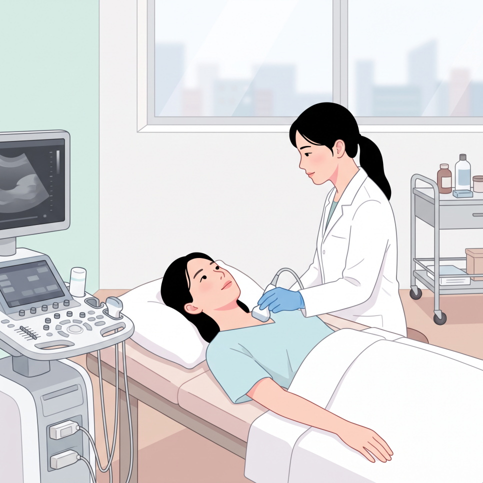
    A painless lump is more likely to be a chronic cyst or lipoma, but in rare cases further testing is necessary.
    That’s why confirming the diagnosis with an ultrasound exam is a safer choice than self-diagnosis.


    How Does Treatment Differ by Cause?

    Knowing the exact cause is key to reducing recurrence and complications.
    Treatment for an armpit lump ranges from simple monitoring to surgical removal, depending entirely on the underlying condition.

    1. Lymphadenitis: May improve with adequate rest and immune care alone; anti-inflammatory pain relievers or antibiotics may be prescribed if needed.
    2. armpit lump related image 5

    3. Sebaceous cyst (epidermal cyst): Avoid squeezing it by hand; having it surgically removed under local anesthesia, including the cyst wall, can lower the risk of recurrence.
    4. armpit lump related image 6

    5. Accessory breast tissue: Since hormones play a major role, periodic monitoring is the standard approach, and surgical removal of the glandular tissue may be considered if pain or discomfort significantly affects daily life.
    Outcome
    Identifying armpit lump causes accurately and managing them appropriately may help some cases avoid major surgery, depending on the underlying condition.
    However, we recommend discussing the diagnosis and treatment plan with a medical professional before making any decisions.

    If you’re concerned about a lump or wound on your skin — not just in the armpit — seeing a specialist is important.
    Burn Treatment Specialists – [Ansan Open Surgery Hospital] Homepage has more information on surgical care.


    Frequently Asked Questions

    1. Is it okay to keep touching an armpit lump to check on it?
    Answer: Bacteria on your hands can cause a secondary infection or worsen inflammation, so it’s best to avoid pressing on or repeatedly touching the lump.

    2. If an armpit lump doesn’t hurt, is it fine to just leave it alone?
    Answer: Even without pain, it could be a chronic cyst, lipoma, or another condition, so it’s safer to monitor any size changes and confirm the diagnosis with an ultrasound exam.

    3. Why does my armpit lump only hurt during my period?

    Answer: It’s likely accessory breast tissue responding to changes in female hormones. This tissue tends to temporarily swell and become painful in sync with the menstrual cycle, then subside afterward.

    4. Which specialist should I see for an armpit lump?
    Answer: Visiting a breast surgeon, general surgeon, or dermatologist allows for an accurate diagnosis using tools like ultrasound, along with guidance tailored to the cause.

    armpit lump related image 7


    References

    · Korea Disease Control and Prevention Agency, "Lymph Node Enlargement," National Health Information Portal. Link
    · Asan Medical Center, "Lymphadenitis," Disease Encyclopedia. Link
    · Asan Medical Center, "Epidermal Cyst," Disease Encyclopedia. Link
    · Health Kyunghyang, "[Accessory Breast Story] Accessory Breast Tissue Is a Real Medical Condition," 2023. Link

    This article is provided for general health information purposes only and is not a substitute for individual diagnosis or treatment. If you have symptoms, please consult a medical professional.

  • Hyperbaric Oxygen Therapy for Burns: Why the 24-48 Hour Golden Window Matters

    Hyperbaric Oxygen Therapy for Burns: Why the 24-48 Hour Golden Window Matters

    SummaryHere’s what you need to know about hyperbaric oxygen therapy for burns: how it works, why the 24-48 hour golden window matters, what the treatment process looks like, and a checklist to help you prepare.

    If a burn from hot water or fire hasn’t even started to scab over after a week,
    it may be more than just a slow recovery.

    In many cases, the underlying issue is reduced blood flow to the damaged tissue, which cuts off its oxygen supply.
    One approach that’s drawn attention for speeding up recovery in these situations is hyperbaric oxygen therapy for burns.

    💡 Quick preview
    This article breaks down how hyperbaric oxygen therapy for burns works, why the golden window matters for recovery, and what to check before starting treatment.
    Read on for the 3 mechanisms that help reduce swelling and lower scarring risk, plus why the 24-48 hour window is so critical.

    hyperbaric oxygen therapy for burns

    What Is Hyperbaric Oxygen Therapy for Burns?

    Hyperbaric oxygen therapy for burns is a treatment in which patients breathe highly concentrated oxygen in an environment with air pressure higher than normal atmospheric pressure, in order to support the recovery of damaged tissue.
    It’s administered inside a sealed piece of equipment called a hyperbaric chamber.

    Under normal breathing conditions, oxygen struggles to reach deep into damaged skin tissue.
    When pressure is increased, however, oxygen dissolves not only into red blood cells but also into blood plasma (serum).

    If this is your first time hearing about the concept, you’re probably wondering how much it actually helps with skin recovery.
    The key idea is that it delivers oxygen directly to injured areas where circulation is compromised.

    hyperbaric oxygen therapy for burns illustration 3

    💡 Key point
    The high-concentration oxygen inhaled under pressure travels through the bloodstream deep into damaged skin, helping to lay the groundwork for cell regeneration.

    hyperbaric oxygen therapy for burns illustration 4


    What Are the Core Mechanisms Behind Faster Recovery?

    Hyperbaric oxygen therapy supports recovery through three main mechanisms: reducing swelling, promoting new blood vessel formation, and supporting white blood cell function.
    When skin tissue is damaged, localized swelling develops and blood flow drops sharply.

    If this state continues, tissue oxygen levels fall further and recovery slows down.
    High-concentration oxygen helps stimulate the formation of new microvessels, allowing fresh blood supply to return to the area.

    hyperbaric oxygen therapy for burns illustration 5

    Elevated blood oxygen levels help reduce swelling
    and support the bactericidal function of white blood cells, which can help guard against secondary infection.

    Together, these combined effects help activate the skin’s regeneration cycle,
    which may in turn help lower the likelihood of scarring.

    🚀 Potential benefits
    ✅ Reduced swelling and improved blood flow at the injury site
    ✅ Support for white blood cell function that may help prevent infection
    ✅ Encouragement of new microvessel and skin cell formation

    Why Does the 24-48 Hour Golden Window Matter So Much?

    The “golden window” in burn care refers to the first 24 to 48 hours after injury, a period that’s understood to help minimize cell death and support the body’s recovery capacity.
    The faster the initial response, the more the extent of damage may be limited.

    hyperbaric oxygen therapy for burns illustration 6

    Prompt, appropriate first aid and care during this period is understood to have a positive effect on outcomes.
    That said, this timeframe reflects general clinical guidance for burn injuries
    and isn’t an absolute standard that applies identically to every patient.

    Missing the golden window raises the risk of deeper tissue damage,
    so it’s advisable to seek a professional evaluation and prompt care immediately after injury.

    ⚠️ A note of caution
    Simply waiting for natural healing without intervention can allow the area of tissue death to expand, so it’s best to get appropriate care within the initial golden window.

    What Does the Hyperbaric Oxygen Treatment Process Look Like?

    Hyperbaric oxygen therapy generally follows this sequence: consultation and evaluation → entering the chamber → progress check.
    Understanding these steps ahead of time can help you receive care safely.

    1. Consultation and evaluation: Medical staff assess the patient’s condition and determine whether the treatment is appropriate.
    2. Entering the chamber: Pressure is raised gradually while the patient breathes high-concentration oxygen for 60 to 90 minutes.
    3. Progress check: Skin recovery is evaluated and the number of remaining sessions is adjusted accordingly.

    hyperbaric oxygen therapy for burns illustration 7

    📋 Checklist
    ✅ Does the facility have equipment that meets recognized safety standards?
    ✅ Is the treatment supervised by qualified medical staff?
    ✅ Were you given clear instructions on equalizing (ear pressure adjustment) before treatment?

    If you’re looking for a specialized hospital that offers hyperbaric oxygen therapy,
    the homepage of Ansan Yeorin Surgery Hospital, a hospital specializing in burn treatment, is a good place to find more information.

    hyperbaric oxygen therapy for burns illustration 8


    Frequently Asked Questions

    1. Is the treatment painful or uncomfortable?
    As pressure increases, you may feel ear fullness similar to what you experience on an airplane. Swallowing or pinching your nose while gently blowing (equalizing) usually resolves this.

    2. How long does each session take, and how many sessions are needed?
    A single session typically takes 60 to 90 minutes, and the total number of sessions varies depending on the severity of the burn and how recovery progresses.

    3. Can it be combined with regular wound dressing?
    Yes. When combined with standard wound care and dressing, it may support skin regeneration alongside those treatments.

    4. Can every burn patient receive this treatment?
    Eligibility depends on the depth and extent of the burn as well as overall health, so it should be decided based on a physician’s evaluation.

    5. Does it completely eliminate scarring after treatment?
    It may help lower the risk of scarring, but results vary by individual and complete scar removal isn’t guaranteed.

    hyperbaric oxygen therapy for burns illustration 9

    hyperbaric oxygen therapy for burns illustration 10

    If you need emergency burn care in the Ansan area, it’s worth checking in advance whether a hospital has dedicated burn specialists and the proper dressing materials on hand.
    Facilities like Yeorin Surgery Hospital, located at 376 Hwarang-ro, Danwon-gu, Ansan-si, Gyeonggi-do, can serve as a reference for the kind of care infrastructure to look for.

    hyperbaric oxygen therapy for burns illustration 11


    References

    · NCBI StatPearls, “Hyperbaric Treatment of Thermal Burns,” StatPearls Publishing, 2023. View source
    · Journal of Burn Care & Research, “Hyperbaric Oxygen Therapy in Burn Care: A Systematic Review of Current Evidence,” Oxford University Press, 2025. View source
    · Doctors News, “Why Is Hyperbaric Oxygen Therapy Needed for Acute Burns?”, Korean Medical Association, 2023. View source


    This article is intended for general health information purposes only and is not a substitute for individual diagnosis or treatment. If you have symptoms, please consult a physician.

  • Flap Necrosis: 3 Causes After Free Flap Surgery (Smoking & Pressure Warning)

    Flap Necrosis: 3 Causes After Free Flap Surgery (Smoking & Pressure Warning)

    SummarySmoking and pressure on the graft site are two of the leading causes of flap necrosis after free flap surgery. Here’s a rundown of reconstructive plastic surgery information — from the basics of vascular anastomosis to a post-surgery care checklist.

    What if a flap that just went through surgery suddenly turned dark red or purple?
    Tissue transferred through free flap surgery is at risk of flap necrosis the moment its blood supply is interrupted.

    Smoking and pressure from improper positioning are two of the most common causes.
    Today, we’ll walk through how free flap surgery works and how to help prevent flap necrosis after the procedure.

    flap necrosis

    Key Points
    ✔ Free flap surgery is a microsurgical reconstruction procedure that transplants tissue along with its blood vessels.
    ✔ Inadequate post-surgery care can lead to flap necrosis.
    ✔ Smoking and pressure are two of the leading risk factors for flap necrosis.

    What Is Free Flap Surgery?

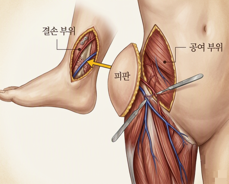
    To put it simply, free flap surgery is a microsurgical reconstruction procedure that removes a whole section of a patient’s own tissue — including its blood vessels — and transplants it to a damaged area.

    flap necrosis illustration 2

    In some cases, a simple skin graft isn’t enough for full recovery.
    This is especially true when the tissue damage is deep or blood circulation to the area is compromised.

    In these situations, surgeons take skin, fat, and even arteries and veins together from a donor site such as the thigh, back, or abdomen.
    But that’s not the end of the process.

    flap necrosis illustration 3

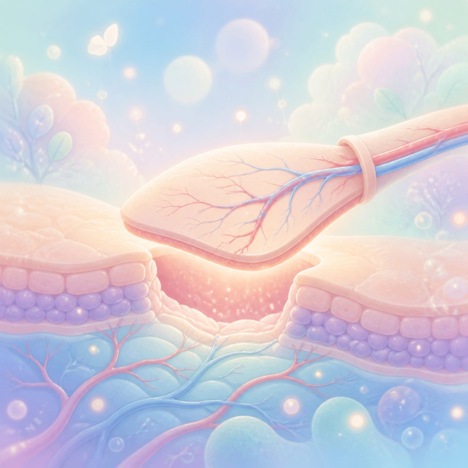
    The tiny blood vessels in the removed tissue must be connected, one by one, to the blood vessels at the recipient site under a microscope.
    This vascular anastomosis process is the core technique of free flap surgery.

    That’s because tissue needs a continuous supply of oxygen and nutrients to survive.
    According to related research, this microvascular connection is considered a critical factor in determining whether the surgery succeeds.

    flap necrosis illustration 4


    Why Does Flap Necrosis Happen? The Effects of Smoking and Pressure

    In short, flap necrosis occurs when the oxygen and nutrient supply to transplanted tissue is cut off, causing the cells to die.

    Nicotine in cigarettes causes blood vessels to constrict.
    Even a single cigarette can narrow the microvasculature.
    That’s why smoking after surgery is considered one of the leading risk factors for flap necrosis.

    Pressure has a similar effect.
    Something as simple as an unconscious sleeping position can cut off blood flow to the grafted area.
    When a blood vessel is compressed, blood clots can form more easily, which can ultimately lead to tissue necrosis.

    ⚠ Precautions
    · Maintain a position that keeps pressure off the surgical site at all times.
    · Avoid smoking, since it constricts blood vessels.
    · Keep warm, as a drop in body temperature can slow blood circulation.
    Risk Factor Effect on the Flap
    Smoking Vasoconstriction, reduced microcirculation
    Pressure/positioning Vessel compression, increased risk of clot formation
    Hypothermia Reduced blood circulation

    An international meta-analysis found that smoking significantly increased the rates of hematoma and overall complications after head and neck cancer reconstruction surgery.
    Another study reported that free flap perfusion tended to be lower in smokers than in non-smokers.

    That said, having a smoking history doesn’t automatically mean flap necrosis will occur.
    Still, since the risk is elevated, extra caution is warranted.

    flap necrosis illustration 5


    When and How Is Free Flap Surgery Performed?

    In short, free flap surgery is used when extensive tissue loss needs to be structurally restored.
    It’s commonly used to fill defects left after severe burns, head and neck cancer surgery, or breast cancer surgery.
    It’s also widely applied for serious skin damage caused by traffic accidents or diabetic foot conditions.

    Checklist — Steps in Free Flap Surgery
    ✅ Step 1: Measure the defect and select the donor site (where tissue will be harvested)
    ✅ Step 2: Harvest the flap, including its blood vessels, from the donor site
    ✅ Step 3: Perform vascular anastomosis (vessel connection) using a surgical microscope
    ✅ Step 4: Close the recipient site and the donor site

    Because this microsurgical technique involves connecting blood vessels with sutures thinner than a human hair, the procedure takes a long time and requires a high degree of precision.


    What Should You Manage After Surgery?

    To put it simply, the key to post-surgery care is keeping the transplanted blood vessels open to prevent flap necrosis.

    flap necrosis illustration 6

    After surgery, blood flow to the transplanted tissue must be monitored continuously.
    If a vessel becomes blocked or a clot forms, the transplanted tissue is unlikely to survive.

    Close monitoring takes place during the first several days.
    Once the tissue has stabilized, patients gradually move into a more routine recovery phase.

    When recovery is managed well,
    it can help support stable flap survival and normal blood flow, and may also help lower the risk of tissue necrosis.

    If you’d like to learn more about tissue defects caused by burns and reconstructive treatment options, visiting Burn Treatment, [Ansan Open Surgery Hospital] Home may also be helpful.

    flap necrosis illustration 7


    Frequently Asked Questions

    1. Can tissue from another person be transplanted instead?
    No — to prevent immune rejection, the general rule is to use only the patient’s own tissue.

    2. Is the donor site (where tissue is harvested) affected afterward?
    Surgeons choose a site that has minimal impact on bodily function and where scarring is less visible, and the donor site is closed with careful surgical technique as well.

    3. How long does recovery take after surgery?
    Hospital stays typically last about one to two weeks, but several months of follow-up monitoring are usually needed before the graft site stabilizes and the patient returns to normal daily activities.

    4. Can the skin color or texture differ from the surrounding area?
    Depending on the donor site used, there can be differences in texture or color, and additional cosmetic procedures may be used to correct this later if needed.

    5. Can smokers undergo free flap surgery?
    Smoking doesn’t necessarily rule out surgery, but because it raises the risk of flap necrosis, quitting smoking before and after surgery is strongly recommended.


    References

    · Asan Medical Center, “Tissue Reconstruction Through Flap Surgery,” Disease Encyclopedia, Asan Medical Center. View source
    · Chang et al., “The impact of smoking on surgical complications after head and neck reconstructive surgery with a free vascularised tissue flap: a systematic review and meta-analysis,” Journal of Plastic, Reconstructive & Aesthetic Surgery (ScienceDirect), 2020. View source
    · “Smoking and microvascular free flap perfusion in head and neck reconstruction: radial free forearm flaps and anterolateral thigh flaps,” Scientific Reports (Nature), 2022. View source
    · Liu et al., “Analysis of the Risk Factors for Free Flap Necrosis in Soft Tissue Reconstruction of the Lower Limbs,” Orthopaedic Surgery (Wiley), 2023. View source

    This article is intended for general health information purposes only and is not a substitute for individual diagnosis or treatment. If you have symptoms, please consult a medical professional.